Orbital Fractures
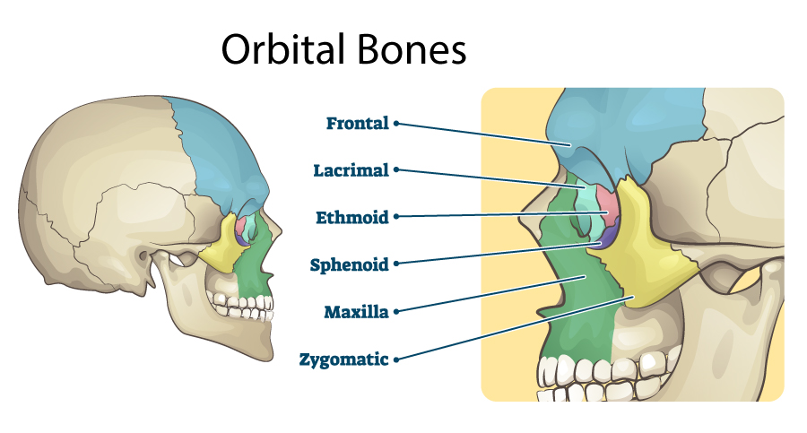
After trauma to the face and eye area (such as after a car accident, fall, assault), one or more bones of the orbit (eye socket) may be broken. This is most commonly diagnosed when a CT scan of the orbits is performed. An associated injury of the eye itself should always be checked for by having an ophthalmologist perform a thorough eye exam. The most commonly fractured bones are the orbital floor (bone below the eye) and the medial wall (bone between the eye and nose). Luckily, many fractures do not need to be fixed and can be observed by an experienced surgeon. Large fractures and fractures resulting in significant double vision often do best if fixed within a few weeks of the injury. Surgery involves placing a synthetic implant over the broken bone to re-establish orbital volume. The incision is most often made within the lower eyelid and results in no visible scar. Board-certified ophthalmologist, fellowship-trained oculoplastic surgeon Katherine J. Zamecki-Vedder, MD, FACS is very experienced in examining and treating patients with orbital fractures.
Get Started
Evaluation and treatment of orbital fractures is often covered by insurance. Call 203-791-2020 to schedule your fracture evaluation with Katherine J. Zamecki-Vedder, MD, FACS.
